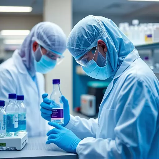
Deep Learning is transforming histopathological image analysis and lab work in Cincinnati, offering unprecedented precision and efficiency in diagnosing complex diseases, especially cancers. By leveraging advanced AI models, researchers streamline cancer diagnostics through liquid biopsies, analyzing circulating tumor cells and nucleic acids in blood samples for real-time results. This revolution promises faster detection, improved accuracy, and personalized treatment planning, enhancing patient outcomes and reducing pathologists' workload in Cincinnati and beyond.
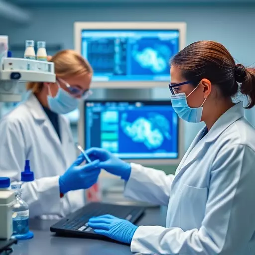
Deep learning is revolutionizing histopathological image analysis, driving significant advancements in medical diagnosis. This article explores the transformative power of deep learning, highlighting its impact on laboratory practices, particularly in Cincinnati’s forward-thinking labs. We delve into how this technology enhances image interpretation, accelerates real-time reporting through liquid biopsy, and improves cancer detection and personalized treatment. By understanding these innovations, we can appreciate the profound effects of deep learning on clinical practice and patient outcomes.
- Deep Learning: Unlocking Precision in Histopathology
- Cincinnati's Lab Revolution: Integrating Deep Learning for Enhanced Image Analysis
- Real-Time Reporting with Liquid Biopsy: Advancing Cancer Diagnosis
- Overcoming Challenges: Data Preparation and Model Training
- Impact on Clinical Practice: Early Detection and Personalized Treatment
Deep Learning: Unlocking Precision in Histopathology
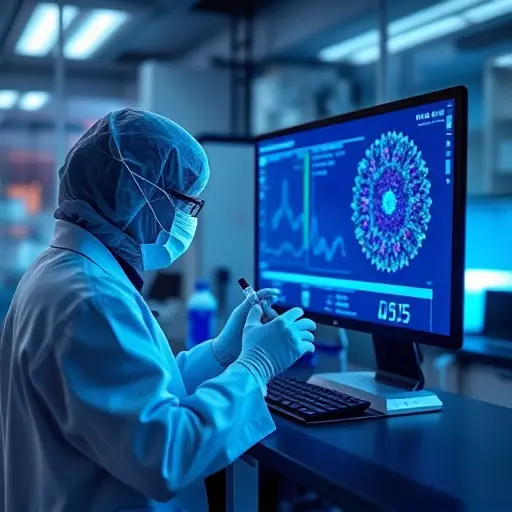
Deep Learning is revolutionizing histopathological image analysis, offering unprecedented precision and efficiency in diagnosing diseases, especially in complex cases that require expert interpretation. This cutting-edge technology mimics the human brain’s neural networks to process and interpret visual data, transforming traditional lab work in Cincinnati and beyond. By analyzing vast amounts of histopathology images at lightning speed, deep learning algorithms can detect subtle patterns and anomalies, enhancing diagnostic accuracy.
In the realm of cancer diagnostics, advances in real-time lab result reporting are made possible through liquid biopsy techniques, which involve analyzing circulating tumor cells and nucleic acids in blood samples. Deep learning plays a pivotal role here by enabling the automated classification and staging of cancer types, providing faster and more accessible patient evaluations. This technology promises to improve patient outcomes and streamline clinical workflows, making it an exciting game-changer in healthcare, particularly when combined with cutting-edge liquid biopsy technologies.
Cincinnati's Lab Revolution: Integrating Deep Learning for Enhanced Image Analysis
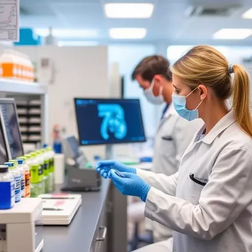
In Cincinnati, a revolution is underway in laboratory work, driven by the integration of deep learning techniques for enhanced histopathological image analysis. This city’s research institutions and healthcare facilities are at the forefront of leveraging advanced artificial intelligence (AI) models to streamline and improve the accuracy of cancer diagnostics. By applying deep learning algorithms to digitalized microscope slides, researchers can automatically identify and classify cellular abnormalities with unprecedented precision.
This breakthrough promises to transform how liquid biopsies are analyzed, significantly speeding up the process of detecting and diagnosing cancer. With real-time lab result reporting becoming a possibility, patients in Cincinnati and beyond can expect more prompt treatment options. The application of deep learning in this context not only enhances diagnostic capabilities but also reduces the workload on pathologists, ensuring that even complex cases receive thorough and timely evaluations.
Real-Time Reporting with Liquid Biopsy: Advancing Cancer Diagnosis
In the realm of cancer diagnosis, advancements in real-time lab result reporting through liquid biopsy are revolutionizing patient care at labs in Cincinnati and beyond. This minimally invasive procedure allows for the analysis of tiny amounts of tumor DNA circulating in the blood, providing a rapid and efficient method to detect and monitor cancer progression. By enabling doctors to access dynamic changes in tumor genetics, liquid biopsy offers significant advantages over traditional histopathological methods, which often rely on fixed tissue samples.
The transformative power of liquid biopsy lies in its ability to deliver real-time reporting, accelerating the diagnostic process and empowering healthcare professionals to make informed decisions promptly. This innovation is particularly impactful for patients with early-stage cancer or those requiring continuous monitoring during treatment. By seamlessly integrating liquid biopsy into lab work in Cincinnati, medical professionals are leveraging cutting-edge technology to enhance cancer diagnostics, ultimately improving patient outcomes and navigating the complex landscape of personalized medicine.
Overcoming Challenges: Data Preparation and Model Training

In the realm of histopathological image analysis, deep learning offers revolutionary advancements, transforming traditional lab work in Cincinnati and beyond. However, harnessing its full potential comes with challenges, especially in data preparation and model training. The complexity of histopathological images requires meticulous pre-processing to enhance features and reduce noise, mirroring the intricate dance of navigating a bustling laboratory. Researchers must carefully curate datasets, ensuring diverse representations of various pathologies, akin to assembling a symphony of different instruments for a harmonious composition.
Advances in real-time lab result reporting are closely tied to these efforts. By optimizing data preparation techniques and employing sophisticated training algorithms, deep learning models can learn nuanced patterns from liquid biopsy samples, revolutionizing cancer diagnostics. This breakthrough promises faster, more accurate detection and classification of diseases, giving patients a significant edge in their fight against the silent enemy within.
Impact on Clinical Practice: Early Detection and Personalized Treatment

The role of deep learning in histopathological image analysis has brought about significant advancements in clinical practice, particularly in early cancer detection and personalized treatment approaches. Through sophisticated algorithms, these models can analyze vast amounts of microscopic images from lab work in Cincinnati more efficiently than traditional methods. This enables healthcare professionals to identify subtle patterns indicative of cancerous cells or precocious conditions at an earlier stage.
Furthermore, advances in real-time lab result reporting are made possible by deep learning integration. Liquid biopsy techniques, for instance, transform cancer diagnostics by analyzing free-floating tumor DNA in blood samples, leveraging deep learning algorithms to interpret these complex data and provide faster, more accurate results. This not only expedites diagnosis but also facilitates personalized treatment planning, where targeted therapies can be tailored to the unique genetic profile of each patient’s tumor.