Lab work in Toledo is at the forefront of Alzheimer's Disease (AD) research, focusing on amyloid beta and tau proteins through advanced testing methods. Similar to tumor gene profiling in targeted cancer therapy, these techniques offer critical insights for AD diagnosis and prognosis, enabling interventions that may slow disease progression. Cytology, a non-invasive technique, assists in early AD detection by analyzing cell samples from bodily fluids or tissues, complementing its role in oncology where it identifies precancerous cells. Together, these innovations navigate the complex landscape of AD care, promising improved patient outcomes through targeted therapies and earlier interventions.
Alzheimer’s Disease (AD) is a devastating neurodegenerative condition with complex pathophysiology. The accumulation of amyloid beta (Aβ) plaques and tau tangles is considered a hallmark of the disease. This article explores the crucial role of testing these proteins in diagnosing AD. We delve into innovative lab work in Toledo, highlighting its impact on research. Additionally, we discuss tumor gene profiling as a potential analogue for targeted therapy in neurology, and examine how cytology aids in detecting precancerous cells, offering insights into early AD diagnosis through similar principles.
- Understanding Alzheimer's Disease: The Importance of Amyloid Beta and Tau Proteins
- Testing Methods: Lab Work in Toledo and its Impact on Alzheimer's Research
- Tumor Gene Profiling: Unlocking Targeted Cancer Therapy and Its Analogies in Neurodegenerative Diseases
- Cytology's Role: Detecting Precancerous Cells and Its Relevance to Early Alzheimer's Diagnosis
Understanding Alzheimer's Disease: The Importance of Amyloid Beta and Tau Proteins

Alzheimer’s Disease (AD) is a progressive neurodegenerative disorder that affects millions worldwide, primarily impacting memory and cognitive functions. Understanding the intricate biological mechanisms behind AD is crucial for developing effective diagnostic tools and treatment strategies. At the heart of this understanding lie two key proteins: amyloid beta (Aβ) and tau.
The accumulation of abnormal Aβ plaques and hyperphosphorylated tau aggregates within the brain are considered hallmark features of AD. These protein deposits lead to neuronal damage, inflammation, and eventual cell death, contributing to memory loss and cognitive decline. Lab work in Toledo, Ohio, and beyond has been instrumental in unraveling these biological processes. Similar to how tumor gene profiling guides targeted cancer therapy, studying Aβ and tau proteins offers insights into the complex pathology of AD, enabling researchers to design targeted interventions. Cytology techniques also play a role in detecting precancerous cells, and similarly, in AD research, they assist in analyzing cerebral fluid and tissue samples for evidence of these pathogenic proteins, thus facilitating earlier diagnosis and intervention.
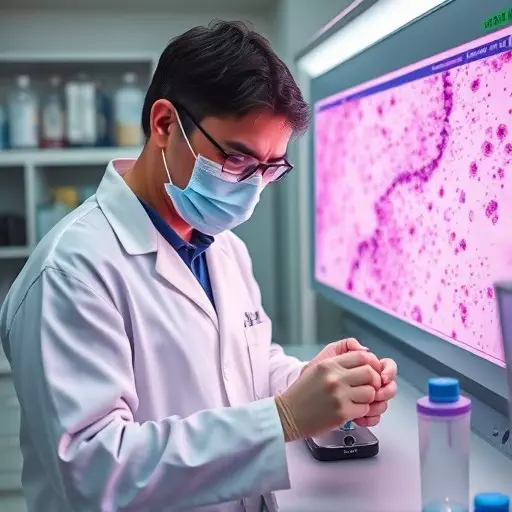
Testing Methods: Lab Work in Toledo and its Impact on Alzheimer's Research

In the advanced realm of Alzheimer’s disease research, lab work in Toledo stands as a beacon, offering sophisticated testing methods that have significantly enhanced our understanding of this complex pathology. One such method involves the analysis of amyloid beta and tau proteins, which play pivotal roles in the progression of the disease. These proteins, when abnormal, aggregate and form distinctive plaques and tangles, hallmarks of Alzheimer’s. By employing cutting-edge techniques, researchers in Toledo are able to detect these protein aggregates with remarkable accuracy, providing invaluable insights into both diagnosis and prognosis.
The impact of this lab work extends beyond local boundaries, reverberating through the global medical community. Similar to how cytology assists in detecting precancerous cells, enabling targeted cancer therapy, the testing methods developed in Toledo are revolutionizing Alzheimer’s research. By profiling these specific proteins, scientists can now identify individuals at risk and develop targeted interventions, mirroring the role of tumor gene profiling in personalized cancer care. This innovative approach promises not just to slow down the disease but also to open new avenues for treatment, reflecting the broader impact of high-quality lab work in advancing medical science.
Tumor Gene Profiling: Unlocking Targeted Cancer Therapy and Its Analogies in Neurodegenerative Diseases

Tumor gene profiling has emerged as a powerful tool in the field of oncology, revolutionizing the way we approach cancer treatment. This advanced lab work in Toledo and beyond involves analyzing a tumor’s genetic makeup to identify specific mutations and alterations that drive cancer growth. By understanding these unique genetic signatures, doctors can tailor targeted therapies aimed at specific molecules or pathways involved in cancer progression. The concept is akin to recognizing the distinct patterns of amyloid beta and tau proteins in Alzheimer’s disease. Just as cytology assists in detecting precancerous cells through microscopic examination, advanced neurological tests identify abnormal protein accumulations that characterize neurodegenerative diseases.
In both cases, early detection plays a crucial role. In cancer, identifying genetic anomalies allows for precise interventions before the disease spreads. Similarly, in Alzheimer’s, understanding the presence and behavior of amyloid beta and tau proteins can lead to more effective treatments. This analogy highlights the value of specialized lab work in Toledo and globally, showcasing how advancements in one field can inform and enhance another, ultimately improving patient outcomes.
Cytology's Role: Detecting Precancerous Cells and Its Relevance to Early Alzheimer's Diagnosis

Cytology plays a pivotal role in early Alzheimer’s disease (AD) diagnosis by assisting in the detection of precancerous cells, although its primary function is often associated with oncology. Similar to its application in targeted cancer therapy through tumor gene profiling in lab work done in Toledo and beyond, cytology helps uncover subtle changes in brain cells that could indicate the initial stages of AD. This non-invasive technique examines cell samples from bodily fluids or tissues, allowing for the identification of abnormalities that might be missed through traditional imaging methods.
By analyzing these cellular remnants, researchers can gain valuable insights into potential neurodegeneration processes. The early detection of such changes is crucial as it enables timely intervention and could potentially slow down or even halt the progression of Alzheimer’s disease before it becomes severe. Thus, cytology’s ability to assist in detecting precancerous cells is not only relevant for cancer management but also holds immense significance in navigating the complex landscape of AD diagnosis and treatment.